The Clitoris Was Finally Fully Mapped in 2026 — 30 Years After the Penis
The Clitoris Was Finally Fully Mapped in 2026 — And Science Is Just Getting Started | thebiologyislove.com
🔥 Breaking — March 2026Amsterdam UniversityNeuroanatomy
The Clitoris Was Finally Fully Mapped in 2026 — And Science Is Just Getting Started
Thirty years after mapping the penis, scientists at Amsterdam University created the world’s first complete 3D nerve map of the clitoris. Here is the full biology, the shocking history of neglect, and why this discovery matters for every woman on the planet.
In March 2026, a team of researchers led by Dr. Ju Young Lee, a neuroanatomist and postdoctoral researcher at Amsterdam University Medical Center, published the world’s first complete three-dimensional nerve map of the human clitoris. The findings were posted on the preprint server bioRxiv on March 20, 2026, and rapidly covered by BBC Science Focus, Smithsonian Magazine, and science outlets worldwide.
The team used synchrotron CT scanning — an advanced X-ray imaging technique powerful enough to resolve structures down to one thousandth of a millimetre — to trace every branch of the main sensory nerve of the clitoris with unprecedented precision. This is 25,000 times smaller than an inch.
💬 Dr. Ju Young Lee — Lead Researcher
“Knowing the anatomy is a prerequisite for understanding the function. Now that we have a better idea about the anatomy, we can start asking more questions about its physiology. That’s why the study is exciting for me as a scientist.” — BBC Science Focus, April 2026
The study examined the pelvises of two postmenopausal women who had donated their bodies to Amsterdam University after death. While the small sample size is a limitation the researchers acknowledge, the structural detail achieved has never before been possible with traditional dissection or clinical imaging.
⚠️ Context — Why a Preprint?
The study was published on bioRxiv as a preprint — meaning it has not yet completed full peer review. This is standard practice for breaking science today; the research is transparent and available, but formal journal peer review is ongoing. The underlying imaging data and methodology have been praised by independent experts.
02 — Biology
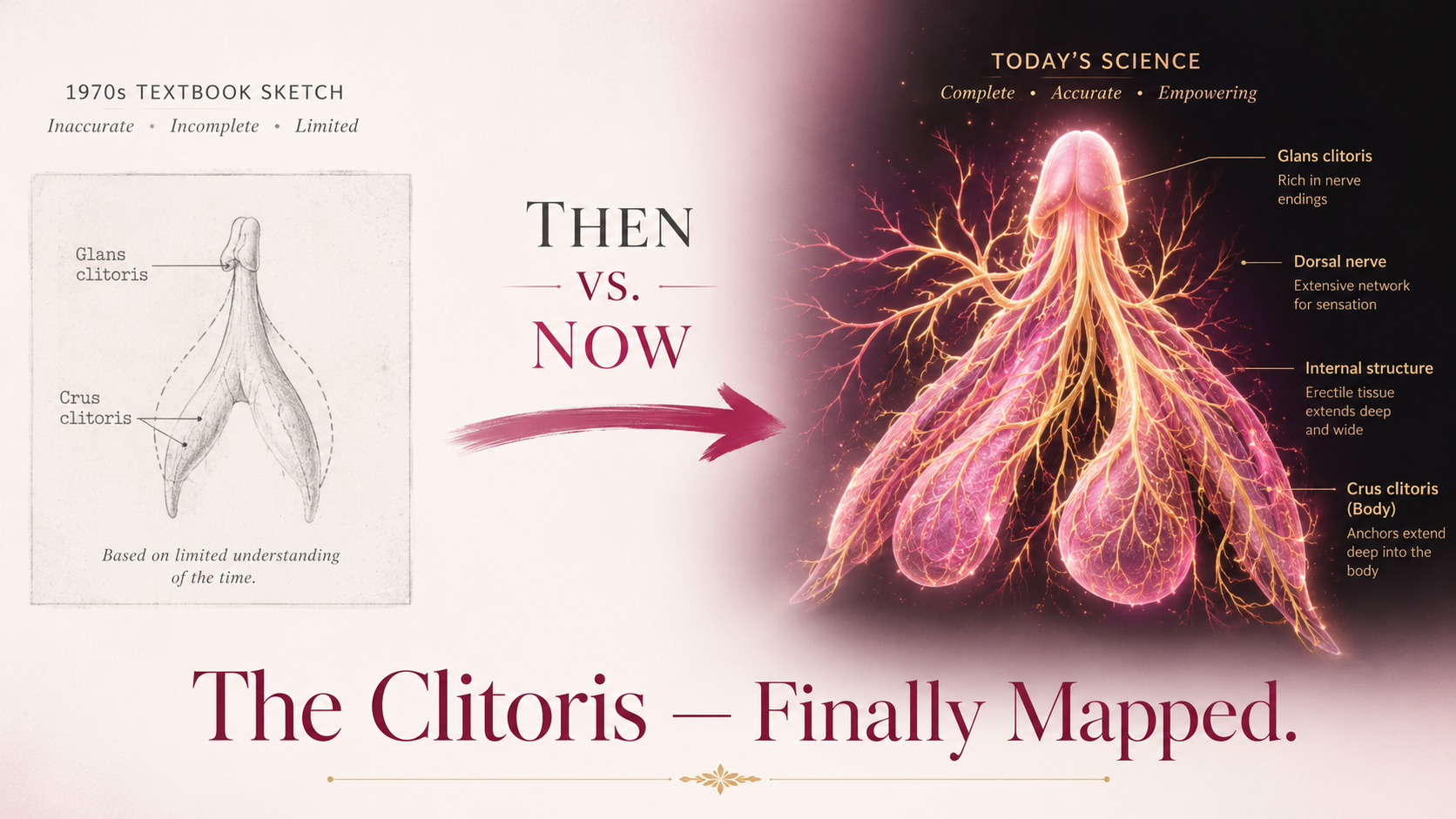
Full Anatomy of the Clitoris — It Is Much Bigger Than You Were Taught
Here is perhaps the single most shocking fact in modern anatomy: only about 10% of the clitoris is visible from outside the body. The remaining 90% is internal — a complex, wishbone-shaped structure tucked within the pelvis. For most of medical history, textbooks drew only the external tip and called it a day.
🔵
Glans Clitoridis
The only externally visible part. Highly sensitive, packed with nerve endings — over 10,000 nerve fibres in a structure roughly the size of a pea. The “tip of the iceberg.”
🔷
Clitoral Body (Shaft)
Extends internally from the glans. Contains erectile tissue (corpus cavernosum) that engorges with blood during arousal — exactly like the penile shaft.
🦋
Crura (Two “Legs”)
Two long arms of erectile tissue that extend inward, wrapping around the vaginal opening. Together they can extend up to 9 cm — larger than an unaroused penis.
💧
Vestibular Bulbs
Two bulbs of erectile tissue on either side of the vaginal opening. When engorged, they press against the vaginal walls — a fact that reshapes our understanding of female sexual response entirely.
🧤
Clitoral Hood (Prepuce)
A fold of skin that covers and protects the glans — the anatomical equivalent of the foreskin. The new nerve map shows nerves extending all the way up into this structure.
⚡
Dorsal Nerve of the Clitoris (DNC)
The main sensory nerve — the star of the 2026 study. Branches like a tree as it reaches the glans. Now fully mapped for the first time in history.
🔬 Size Reality Check
The full clitoris, when aroused, extends up to 9 centimetres internally. Medical textbooks for decades showed a structure roughly 1–2 cm long. Research now confirms it is at least twice as large as depicted — and its nerve density is up to 15 times greater than that of the penis.
💡 Mnemonic — Remember the Parts of the Clitoris
“Great Biologists Can Visit Daring Nerves”
Glans — the visible tip Body (shaft) — internal erectile tissue Crura — the two “legs” extending inward Vestibular bulbs — flanking the vaginal opening Dorsal nerve of the clitoris — main sensory nerve Nerve hood (prepuce/clitoral hood) — the protective cover
03 — The Nerve Map
What the 3D Nerve Map Actually Revealed
The centrepiece of the 2026 study is the first complete map of the Dorsal Nerve of the Clitoris (DNC) — the primary sensory nerve responsible for clitoral sensation. Here is what the map revealed that was not known before:
❌ Old Understanding
Nerve tapers and shrinks as it reaches the glans
→
✅ New Discovery (2026)
Nerve branches intricately — like a tree — throughout the entire glans
❌ Old Understanding
Nerve ends at the glans boundary
→
✅ New Discovery (2026)
Nerve extends upward into the clitoral hood and skin above it
❌ Old Understanding
~8,000 nerve fibres (estimated from animal studies)
→
✅ New Discovery (2022–2026)
10,000+ nerve fibres confirmed in human tissue
How Was It Done? — The Technology
The team used synchrotron phase-contrast CT scanning — a specialised form of X-ray imaging available at only a handful of facilities worldwide. Unlike regular CT scans, this technique passes extremely bright, coherent X-ray beams through tissue, allowing researchers to trace nerve pathways at sub-millimetre resolution without destroying the tissue. The result was a digital 3D reconstruction of every nerve branch — something traditional dissection could never achieve.
🔬 The Technique in Simple Terms
Think of it like Google Maps, but for nerves inside the body. Traditional anatomy was like drawing a map from memory. Synchrotron CT is like having a satellite photograph every street, lane, and alley at once — down to centimetre precision. That is what Dr. Lee’s team did for the clitoris for the very first time.
✅ Expert Reaction
“The team’s results help correct long-standing gaps and misconceptions in our understanding of female sexual anatomy.” — Caroline Pukall, Queen’s University Canada, commenting to Smithsonian Magazine
04 — History of Neglect
A History of Scientific Neglect — How Did This Take So Long?
The 2026 nerve map did not happen in a vacuum. It is the latest chapter in a long, painful story of how female anatomy — particularly the clitoris — was ignored, erased, and misrepresented by medical science for centuries.
1
Pre-1900s
Medical textbooks routinely omit or minimise the clitoris. Female genitalia described primarily in relation to reproduction. One famous 19th-century anatomy text described female genitalia as a “failure” of male development.
2
1976
The widely cited estimate of 8,000 nerve fibres in the clitoris is published — based on research conducted on cows, not humans. This figure enters medical textbooks and persists unchallenged for nearly 50 years.
3
1996
Researchers fully map the nerves of the penis. This data directly informs surgical technique, improving outcomes for prostate surgery, penile reconstruction, and gender-affirming care for trans women.
4
2005
Australian urologist Dr. Helen O’Connell publishes the first full MRI-based map of the clitoris, revealing its true internal size and structure for the first time. She describes how her medical training textbooks never even mentioned the clitoris.
5
2022
Researchers at Oregon Health and Science University count human clitoral nerve fibres from tissue donated by transgender men — confirming over 10,000 fibres, correcting the 1976 cow-based estimate at last.
6
March 2026
Dr. Ju Young Lee’s team at Amsterdam University completes the first 3D nerve map of the clitoris using synchrotron CT — 30 years after the same was done for the penis. Dr. Lee calls it “the starting point of clitoris science.”
😔 The Taboo Factor — In Dr. Lee’s Own Words
“There is a societal taboo attached to female sexuality. The taboo is an obstacle to conducting scientific investigation. Much more awareness is required, starting with the knowledge that the clitoris is actually quite large.” — Dr. Ju Young Lee, Smithsonian Magazine, 2026
05 — The Research Gap
Clitoris vs Penis — The Numbers That Reveal the Bias
Comparison Point
Penis Research
Clitoris Research
Nerve fibres counted (human)
Well-established for decades
First confirmed in 2022
Full nerve map (3D)
Completed 1996
Completed March 2026
Scientific papers on glans
20× more papers
20× fewer papers
True size in textbooks
Accurately depicted
Shown at ½ actual size for decades
Nerve density vs other
Reference point
Up to 15× denser than penis
Surgical protection protocols
Long established
Now being developed from 2026 data
⚡ The Staggering Stat
There are 20 times more scientific papers on the penile glans than on the clitoral glans. The clitoris exists in roughly half the world’s population. The research gap is not a coincidence — it is a reflection of centuries of gender bias embedded into science itself.
06 — Clinical Significance
Why Does This Nerve Map Matter for Medicine?
This is not purely academic. The 3D nerve map of the clitoris has direct, life-changing implications for surgical practice and patient outcomes across multiple fields of medicine:
🍼
Childbirth & Episiotomy
Episiotomy (surgical incision during childbirth) and perineal tears can damage clitoral nerves. Knowing exactly where nerves run enables surgeons to protect them — reducing long-term sexual dysfunction after birth.
🌈
Gender-Affirming Surgery
Clitoroplasty (for trans women) and phalloplasty (for trans men) both involve clitoral tissue. Accurate nerve maps mean surgeons can preserve sensation and function during these life-affirming procedures.
💛
FGM Reconstruction
For survivors of female genital mutilation, reconstructive surgery can restore partial sensation. This nerve map is a critical guide for surgeons attempting to reconnect severed nerve pathways.
🏥
Pelvic Surgery
Hip replacements, mesh surgery, bladder procedures — many pelvic operations risk nerve damage. Surgeons can now navigate with precision, the same way penis nerve maps transformed prostate surgery outcomes in the 1990s.
🧠
Sexual Dysfunction Research
Conditions like provoked vestibulodynia, clitoral phimosis, and persistent genital arousal disorder all involve clitoral nerves. A detailed map is the foundation for understanding and treating these conditions.
📚
Medical Education
Future anatomy textbooks will finally depict the clitoris accurately — full size, full nerve structure. An entire generation of doctors will be trained on facts, not historical omissions.
🌍 The Bigger Picture
“If you don’t know where the nerves are, you risk damaging them.” — Dr. Maria Uloko, Oregon Health and Science University, 2022. This applies to every surgery performed near the clitoris, for any reason. The 2026 map changes the standard of care permanently.
07 — Sources
📖 Read the Original Sources
Science is only as good as its sources. Here are the key publications and articles behind this discovery — read them directly:
Lead researcher: Dr. Ju Young Lee · Institution: Amsterdam University Medical Center · Technique: Synchrotron phase-contrast CT scanning · Published: March 20, 2026 on bioRxiv · First complete 3D map of the Dorsal Nerve of the Clitoris (DNC) · Resolution: down to 0.001 mm · 30 years after equivalent penis nerve mapping (1996).
🧬 Clitoris Anatomy at a Glance
Parts: Glans · Clitoral body · Crura (2) · Vestibular bulbs (2) · Clitoral hood
Only 10% is visible externally · Full erect length up to 9 cm · 10,000+ nerve fibres · Nerve density up to 15× greater than penis · Main nerve: Dorsal Nerve of the Clitoris (DNC).
⚡ Key Corrections the Map Made
❌ Old: DNC tapers at the glans → ✅ New: DNC branches like a tree throughout the glans
❌ Old: Nerve ends at glans → ✅ New: Nerve extends into the clitoral hood
❌ Old: ~8,000 fibres (cow estimate, 1976) → ✅ New: 10,000+ confirmed in human tissue (2022)
🏥 Why It Matters Clinically
Childbirth / episiotomy · Gender-affirming surgery · FGM reconstruction · Pelvic surgery nerve protection · Sexual dysfunction treatment · Medical education reform.
💡 The Research Gap — Remember This
“20 times more papers on the penis. Half the world has a clitoris.”
There are 20× more scientific publications on the penile glans than the clitoral glans. The penis nerve map was completed in 1996. The clitoris nerve map: 2026. That is a 30-year gap for an organ present in half the global population. Dr. Lee called the 2026 map only the “starting point” — much more remains unknown.
09 — Practice Questions
🧠 Test Yourself — MCQs
2026 Discovery
Q1. Which institution published the first complete 3D nerve map of the clitoris in 2026?
✅ B — Amsterdam University Medical Center. Led by Dr. Ju Young Lee, a postdoctoral neuroanatomist. The team used synchrotron CT scanning to produce the map — posted on bioRxiv on March 20, 2026. Oregon Health and Science University (option C) was responsible for the 2022 nerve fibre count study, a different but related breakthrough.
Anatomy
Q2. Approximately what percentage of the clitoris is visible externally?
✅ D — ~10%. Only the glans clitoridis (the external tip) is visible. The remaining 90% — the body, crura, and vestibular bulbs — is entirely internal. When fully aroused, the complete structure can extend up to 9 cm. Medical textbooks historically depicted only the external glans, dramatically underselling the organ’s true size and complexity.
Neuroanatomy
Q3. What is the name of the main sensory nerve of the clitoris mapped in the 2026 study?
✅ C — Dorsal nerve of the clitoris (DNC). This is the primary sensory nerve supplying the clitoris, technically a branch of the pudendal nerve. The 2026 study showed that rather than tapering at the glans (as previously assumed), the DNC branches intricately like a tree throughout the glans, and extends further into the clitoral hood than anyone had mapped before.
History
Q4. The original 1976 estimate of clitoral nerve fibres (~8,000) was based on research conducted on:
✅ C — Cows. The 1976 figure of ~8,000 nerve fibres entered medical literature based on bovine (cow) anatomy, not human. This estimate was cited in textbooks for nearly 50 years without being verified in human tissue. It took until 2022 for researchers at Oregon Health and Science University to count human clitoral nerve fibres directly — confirming over 10,000, correcting a half-century of animal-derived misinformation.
Clinical Significance
Q5. How does the nerve density of the clitoris compare to that of the penis?
✅ C — Up to 15 times greater. Despite being largely overlooked by medical science, the clitoris packs an extraordinary nerve density — up to 15× that of the penis, in a structure concentrated primarily in the glans. This fact alone explains why clitoral nerve damage during surgery has such profound consequences, and why accurate nerve mapping is so critically important for surgical planning.