Table of Contents
The Helix Turn Helix (HTH) motif is present in many prokaryotic and eukaryotic DNA binding proteins. The Helix Turn Helix motif was first DNA-binding structure to be identified.
The structure of Helix Turn Helix motif
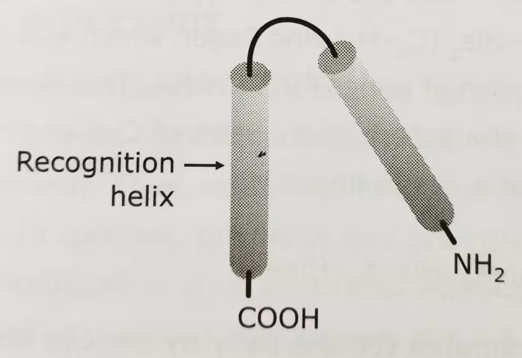
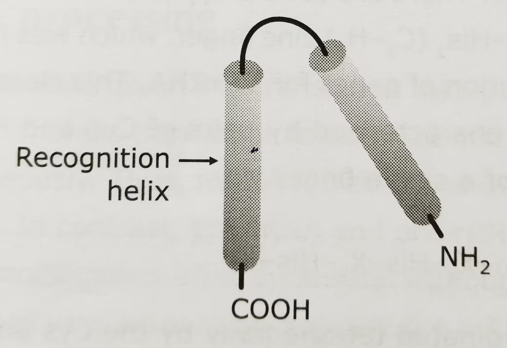
- The motif is made up of two α helices separated by a β turn, made up of four amino acids, the second of which is generally glycine.
- This turn, in conjunction with the first α helix, positions the second α helix on the surface of the protein in an orientation that enables it to fit inside the major groove of a DNA molecule.
- This second α helix is known as recognition helix (C terminal α helix).
- The HTH is generally 20 or so amino acids in length and so is just a small part of the protein as a whole.
The diagrammatic representation of HTH motif:

The homeodomain:
- The homeodomain is an extended HTH motif possessed by proteins and coded by homeotic genes.
- It is made up of the highly conserved domain of 60 amino acids encoded by a 180 bp DNA sequence called a homeobox.
- It has three α helices and helices 2 and 3 are separated by a β turn.
- Helix 3, which is 17 amino acids residues long, acts as a recognition helix.
- Helix 3 binds to the major groove of DNA.
- Helix 1 makes contacts within the minor groove.
Examples:
| Binding motif | Organism | Regulatory protein |
|---|---|---|
| Helix Turn Helix | E. coli | Lac repressor, CAP |
| Helix Turn Helix | Phage | Cro |
Other DNA binding motifs:
- Introduction to DNA binding motifs: https://thebiologyislove.com/dna-binding-motifs/
- helix-loop-helix: https://thebiologyislove.com/helix-loop-helix-motif/
- leucine zipper motif: https://thebiologyislove.com/leucine-zipper-motif/
- zinc finger motif: https://thebiologyislove.com/zinc-finger-motif-c2-h2/
Facebook link: https://www.facebook.com/share/p/7ddyNGS2KjA57CBC/?mibextid=oFDknk
Instagram link: https://www.instagram.com/reel/C8jnR0Wx5B2/?igsh=b3F0bmhnc2ZvNGRm

