
Table of Contents
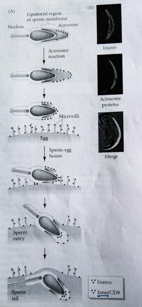
The mechanism of mammalian gamete fusion is still controversial. On the sperm side of the mammalian gamete fusion process, an immunoglobulin-like protein, named Izumo, is originally found in the membrane of the acrosomal granule.
The process of gamete fusion
After the acrosome reaction, Izumo redistributes along the surface of acrosome-reacted sperm, where it is found primarily in the equatorial section (the junction between the inner acrosomal membrane and the sperm cell membrane), where the mammalian sperm-egg binding takes place.
Sperm from mice carrying loss-of-function mutations in the Izumo gene are able to bind and penetrate the zona pellucida but they are not able to fuse with egg cell membrane.
Human sperm also contain Izumo protein and antibodies directed against Izumo prevent sperm-egg fusion in humans as well. There are other candidates for sperm fusion proteins, and there may be several sperm-egg binding systems operating, each of which may be necessary but not sufficient to ensure appropriate gamete binding and fusion.
Izumo-Juno binding:
- Izumo binds to an oocyte protein called Juno, and egg deficient in Juno cannot bind or fuse with acrosome-reacted sperm.
- The interaction of Izumo and Juno recruits the egg membrane protein CD9 to the location of the sperm-egg adhesion.
- This protein appears to be involved with sperm-egg fusion, since female mice with the CD9 gene knocked out are found to be infertile due to the fusion defects.
- But CD9 protein is also known to be critical for the fusion of myocytes (muscle cell precursors) to form striated muscle.
The diagram of gamete fusion in fertilization event in mouse:
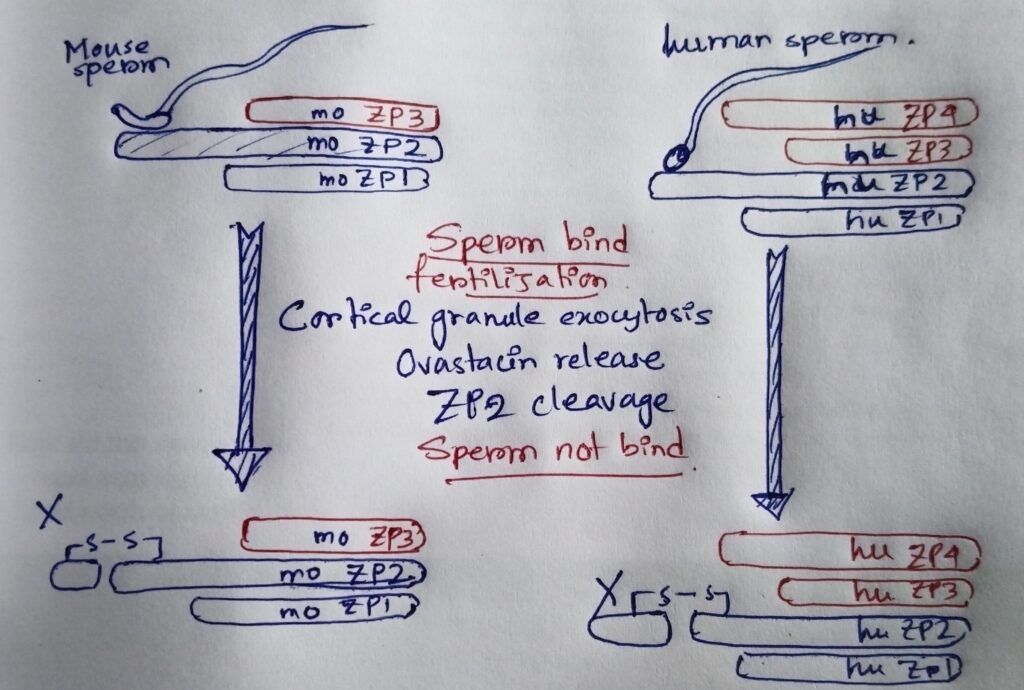
The prevention of polyspermy:
- In mammals, no electrical fast block to polyspermy has yet been observed.
- However, a slow block to polyspermy occurs when enzymes released by the cortical granules modify the zona pellucida sperm receptor proteins such that they can no longer bind to the sperm.
- ZP2 (One of the major glycoproteins of zona pellucida) is clipped by the protease ovastacin and loses its ability to bind sperm.
- Ovastacin is found in the cortical granules of the unfertilized eggs and is released during cortical granule fusion.
- Polyspermy occurs more frequently in mouse eggs bearing mutant ZP2 that cannot be cleaved by ovastacin.
- Another slow block to polyspermy involves the Juno protein.
- As the sperm and egg membranes fuse, Juno protein appears to be released from the plasma membrane. Thus, the docking site for the sperm would be omitted.
- Moreover, this soluble Juno protein can bind perm in perivitelline space between the zona pellucida and oocyte; thereby preventing the sperm from seeing any Juno protein that may still reside on the oocyte membrane.
The diagram of prevention of polyspermy:
Other related notes:
- Cell cycle checkpoints: https://thebiologyislove.com/cell-cycle-checkpoints/
- Patterns of cleavage: https://thebiologyislove.com/patterns-of-cleavage/
Facebook link: https://www.facebook.com/share/p/tqBpuJ3oDWh41veh/?mibextid=oFDknk
The note is made from the Developmental Biology book by Gilbert.