
Table of Contents
Cell junctions are cell-to-cell linkage and cell to the extracellular matrix at specialized contact sites. Cell junctions can be classified into three functional groups- occluding junctions, anchoring junctions and communicating junctions.
Cell Junctions- Occluding Junctions:
Occluding junctions seal cells together in an epithelium in a way that prevents even small molecules from leaking from one side of the sheet to the other (i.e. forms permeability barrier across epithelial cell sheets). These junctions are two types – tight junction and septate junction.
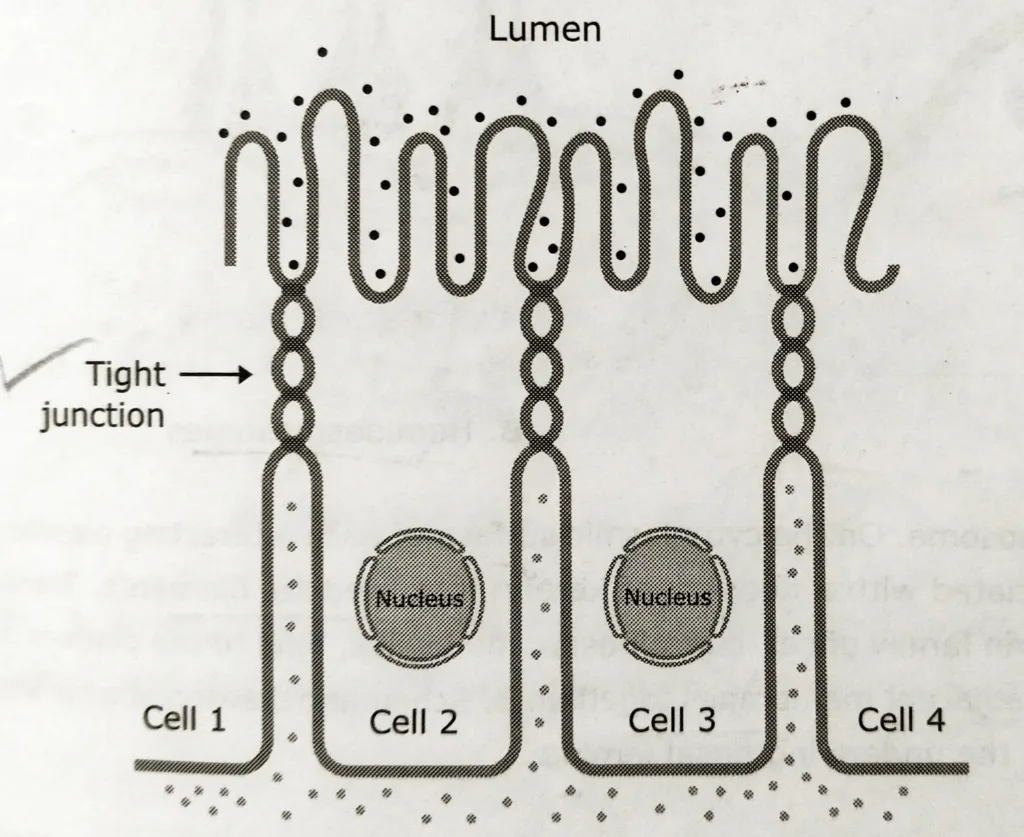
Tight Junction (Zonula occludens)
These are cell-cell occluding junctions mediated by two major transmembrane proteins -claudins and occludin.
Claudins and occludin are associated with intracellular peripheral membrane proteins called ZO proteins. Tight junctions make the closest contact between adjacent cells and prevent the free passage of molecules (including ions) across an epithelial sheet in the spaces between cells. They also maintain the polarity of epithelial cells by preventing the diffusion of molecules between the apical and basolateral regions of the plasma membrane.
Septate junction
These are the main occluding junctions in invertebrates.
Cell Junctions- Anchoring Junctions
Anchoring junctions mechanically attach cells (and their cytoskeletons) to their neighbours or to the extracellular matrix and perform the key task of holding cells together into tissues. It includes two main types of junctions – adherens junction and desmosome.
Adherens Junctions
Adherens junctions connect actin bundles from cell to cell or cell to the extracellular matrix.
Adhesion belt (or zonula adherens) :
It is the cell to cell junction, mediated by the actin filaments and proteins belonging to the cadherin family. Adhesion belts are usually located near the apical surface, just below the tight junctions.
Focal contact (or adhesion plaque):
It is a cell-matrix junction which is mediated by transmembrane adhesion proteins of the integrin family and by actin filament.
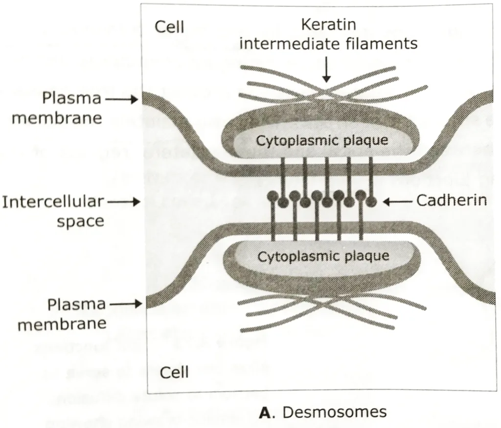
Desmosomes
- Desmosomes are buttons like points of intracellular contacts which bond neighbouring cells together.
- It has a dense cytoplasmic plaque which is composed of a mixture of intracellular attachment proteins, including plakoglobin and desmoplakins.
- The cytoplasmic plaque is responsible for connecting the cytoskeleton to the transmembrane linker proteins of the cadherin family of cell-cell adhesion molecules.
- Desmosomes contain two specialized cadherin proteins, desmoglein and desmocollin. Through extracellular domains, cadherins are responsible for holding the adjacent membranes together.
- Each plaque is associated with a thick network of keratin intermediate filaments (in most epithelial cells) and desmin intermediate filaments ( in heart muscle cells), which are attached to the surface of the plaque.
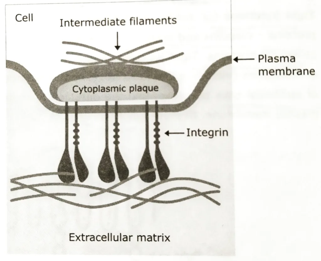
Hemidesmosomes
This is considered as the half desmosomes. These resemble desmosomes type cell junctions, but instead of joining adjacent epithelial cell membranes, they connect the basal surface of epithelial cells to the underlying basal lamina – a specialised mat of extracellular matrix at the interface between the epithelium and connective tissue. The transmembrane linker proteins in hemidesmosomes belong to the integrin family of extracellular matrix receptors, rather than to the cadherin family of cell -cell adhesion proteins used in desmosomes.
Characteristics of Anchoring Junctions
| Cell Junctions types | Transmembrane Protein | Intracellular linkage |
| Zonula Adherens | Cadherin | Actin filaments |
| Focal Contact | Integrin | Actin filaments |
| Desmosome | Cadherin | Intermediate filaments |
| Hemi-desmosome | Integrin | Intermediate filaments |
Cell junctions- Communicating Junctions
Communicating Junctions mediate the passage of chemical or electrical signals from one interacting cell to its partner.
Gap Junctions
- Gap junctions serve as direct connections between the cytoplasm of adjacent cells.
- They act as channels, which allow inorganic ions and other small water-soluble molecules upto 1000 Da to pass directly from the cytoplasm of one cell to the cytoplasm of another cell, thereby coupling the cells both electrically and metabolically.
- In nervous tissue, some neurons are connected by gap junctions through which ions pass rapidly, thereby allowing very rapid transmission of electrical signals.
- Gap junctions also allow the passage of some intracellular signaling molecules, such as cAMP and calcium, between adjacent cells, potentially coordinating the responses of cells in tissues.
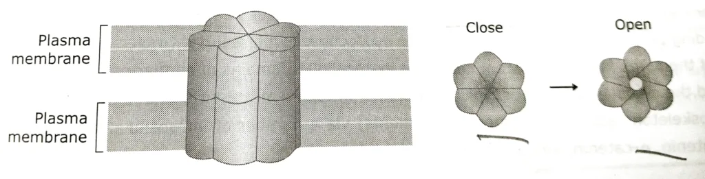
- Gap junction channels are unusual in that they typically remain open at rest, and only close under specific conditions. They close in response to low pH, high intracellular calcium ions concentration and voltage difference between the cells.
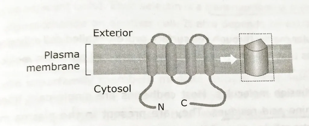
- Gap junctions consist of transmembrane proteins called connexins. All connexins have four transmembrane helices.
- Six connexins assemble to form a cylinder with an open aqueous pore in its center called connexon.
When the connexons in the plasma membranes of two cells in contact are aligned, they form a continuous aqueous channel called gap junction. - A completely different family of proteins, the innexins from the gap junction in invertebrates.
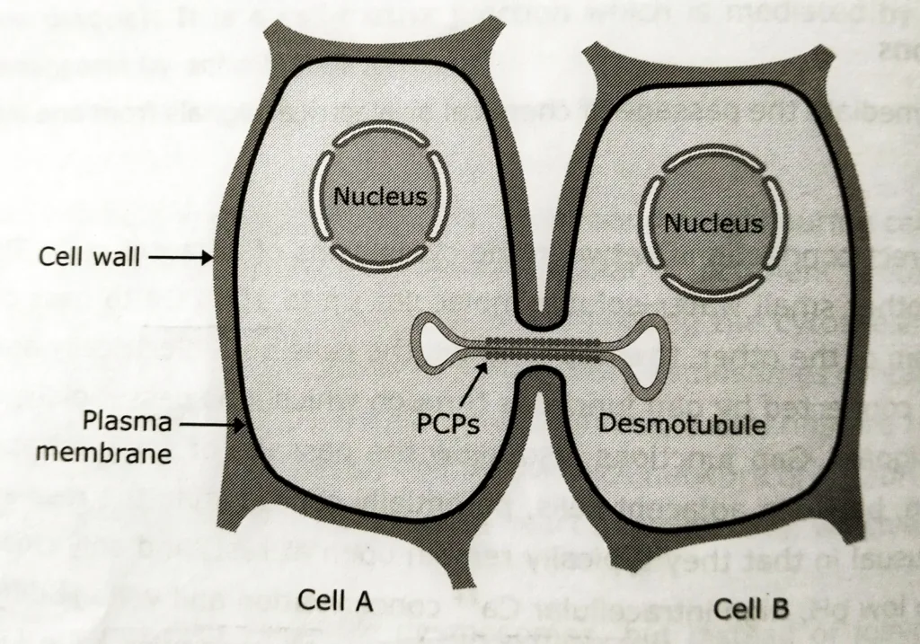
Plasmodesmata
- Adjacent plant cells communicate with each other through cytoplasmic connections. These types of cell junctions are called plasmodesmata (singular- plasmodesma), which function analogously to animal cell gap junctions.
- Despite their similarities in function, plasmodesmata are structurally unrelated to the gap junctions.
- At each plasmodesma, the plasma membrane of one cell is continuous with that of its neighbor, generating an open channel between the two cytosols.
- An extension of the smooth endoplasmic reticulum (SER) called desmotubule passes through the pore, leaving a ring of surrounding cytoplasm through which ions and small molecules are able to pass freely between the cells.
Other related notes:
- Cell cycle checkpoints: https://thebiologyislove.com/cell-cycle-checkpoints/
- Nondisjunction and Aneuploidy: https://thebiologyislove.com/nondisjunction-and-aneuploidy/
- RAS-MAP kinase pathway: https://thebiologyislove.com/ras-map-kinase-pathway/
Facebook link: https://www.facebook.com/share/p/qL7pXZ2Qmy9sxqKE/?mibextid=oFDknk
Instagram Link: https://www.instagram.com/p/C7mQv5IpAuZ/?igsh=MWlnNHJnY3YzOHV4cQ==