- Why This Chapter Matters
- Cell Cycle — Overview
- Interphase: G₁, S & G₂ Phases
- Mitosis — Equational Division
- Cytokinesis
- Meiosis — Reductional Division
- Prophase I Substages (LZPDD)
- Meiosis II
- Mitosis vs Meiosis — Comparison Table
- Cell Cycle Regulation & Checkpoints
- Significance of Cell Division
- Previous Year MCQs
- Quick Revision
Why This Chapter Matters for Every Exam
Every living organism depends on cell division for growth, repair, and reproduction. Cell cycle and cell division is one of those rare chapters that is simultaneously easy to score in NEET (direct fact-based questions) and deep enough to appear as analytical questions in CSIR NET and GATE.
The Cell Cycle — Life’s Repeating Loop
The cell cycle is the sequence of events a cell undergoes from its formation (after a previous division) to its own division into two daughter cells. It was first described clearly by Howard and Pelc (1953). The average duration for a human cell is about 24 hours.
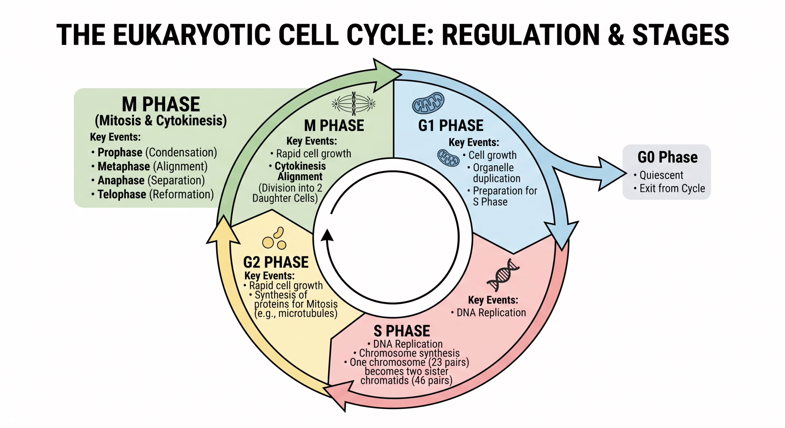
G₀ (quiescent stage) = cells that have exited the cell cycle and are metabolically inactive but alive. Neurons and muscle cells are largely in G₀. Some cells (stem cells, liver cells) can re-enter the cycle when stimulated.
Interphase — The Cell’s Preparation Phase
Interphase is the resting phase only in name — the cell is actually extremely active, growing and preparing for division. It occupies about 90–95% of the total cell cycle time. It consists of three sub-phases: G₁, S, and G₂.
The cell grows in size, synthesises proteins, RNA, and organelles. DNA content = 2C, chromosome number = 2n. The cell prepares for DNA replication. Restriction point (R point) at end of G₁ — after this, cell is committed to division. Cells that are not dividing exit here to G₀. Duration: ~11 hours (longest phase).
DNA replication occurs — each chromosome replicates to form two sister chromatids joined at the centromere. DNA content doubles: 2C → 4C. Chromosome number remains 2n (chromatids joined = not yet separate chromosomes). Centrosome also duplicates here. Duration: ~8 hours.
Continued protein synthesis, growth. Cell checks that DNA replication is complete and accurate. Tubulin protein synthesized for spindle formation. DNA content = 4C, chromosome number = 2n (each as two sister chromatids). Duration: ~4 hours.
Cell exits the cycle — metabolically active but not dividing. Not the same as cell death. Examples: neurons (permanent G₀), liver cells (can re-enter). Senescent cells (aged, non-dividing) are also described as G₀. Important NEET trap: G₀ cells are NOT metabolically inactive — they are functionally active but non-dividing.
Add G₀ = “take a break and Go 0 dividing”
Mitosis — Equational Division (2n → 2n)
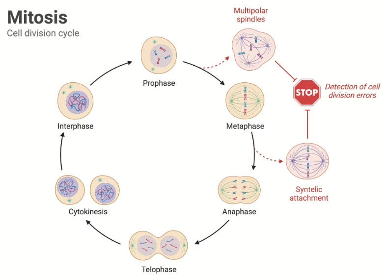
Mitosis is the type of cell division in which a parent cell divides to produce two genetically identical daughter cells, each with the same chromosome number as the parent. It occurs in somatic (body) cells and is responsible for growth, repair, and asexual reproduction. Occurs in 4 stages: Prophase → Metaphase → Anaphase → Telophase.
Add cytokinesis after telophase = “People Meet And Talk, then Celebrate (Cytokinesis)”
Earliest visible stage of M phase.
• Chromatin fibres condense → chromosomes become visible (each = 2 sister chromatids joined at centromere)
• Nucleolus disappears
• Nuclear envelope starts to break down
• Centrosomes move to opposite poles; spindle fibres start forming (asters appear)
• Longest phase of mitosis
Best stage to count chromosomes!
• Nuclear envelope completely disappears
• Chromosomes reach maximum condensation (shortest and thickest)
• Chromosomes align at the metaphase plate (equatorial plate) — midline of cell
• Spindle fibres (from opposite poles) attach to centromeres via kinetochores
• Two kinetochores per chromosome (one per chromatid), each attached to opposite poles
• Centromeres split — sister chromatids separate and are now called chromosomes
• Chromosomes move toward opposite poles pulled by spindle fibres (shortening of kinetochore microtubules)
• Cell elongates (non-kinetochore / polar microtubules push poles apart)
• At this point: 4n chromosomes total (2n at each pole) but still 2C per daughter cell-to-be
• Shortest phase of mitosis
• Cell looks like a butterfly — two groups of chromosomes at opposite poles
• Chromosomes reach poles and begin to decondense
• Nuclear envelope reforms around each set of chromosomes
• Nucleolus reappears
• Spindle fibres disassemble
• Result: two nuclei, each with 2n chromosomes (identical to parent)
Cytokinesis — Division of Cytoplasm
After karyokinesis (nuclear division) is complete, the cytoplasm divides in a process called cytokinesis. This differs between animal and plant cells.
| Feature | Animal Cell | Plant Cell |
|---|---|---|
| Mechanism | Cleavage furrow — constriction of plasma membrane inward | Cell plate formation — vesicles from Golgi fuse at equatorial plate |
| Direction | Outside in (centripetal) | Inside out (centrifugal) |
| Structure formed | No new wall — just plasma membrane pinched | New cell wall formed from cell plate |
| Key protein | Actin-myosin contractile ring | Phragmoplast (microtubule structure) |
| Result | Two daughter cells separated by plasma membrane | Two daughter cells separated by cell plate → new cell wall |
Cytokinesis = division of the cytoplasm.
In some cells, karyokinesis can occur without cytokinesis → syncytium (multinucleate cell, e.g., skeletal muscle fibres, coconut endosperm during early development).
Meiosis — Reductional Division (2n → n)
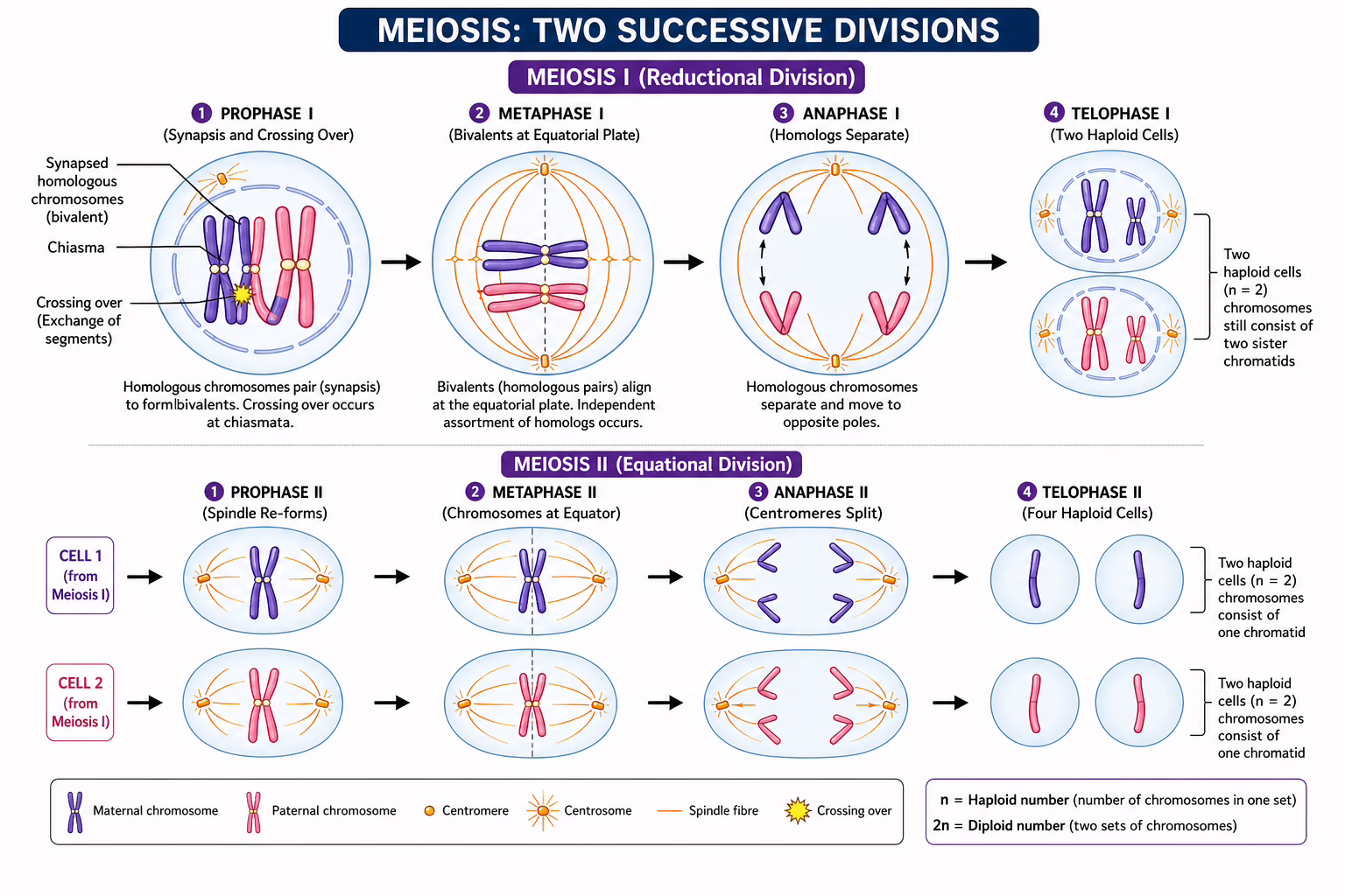
Meiosis is a special type of cell division that produces four haploid daughter cells from a single diploid parent cell. It occurs only in gonads (testes and ovaries) for gamete formation. It involves two successive divisions: Meiosis I (reductional) and Meiosis II (equational).
Meiosis I — The Reductional Division
Meiosis I reduces the chromosome number from diploid to haploid. It is the true reductional division. The most complex and longest phase is Prophase I, which is further divided into 5 substages.
Prophase I — The Most Complex Phase in Biology
Prophase I of Meiosis I is subdivided into 5 stages: Leptotene, Zygotene, Pachytene, Diplotene, and Diakinesis. This is one of the most heavily tested topics in NEET, CSIR NET, and GATE.
Each stage has ONE key event — master that and you’ll answer any NEET question on this topic.
| Substage | Key Event | NEET Trigger Word |
|---|---|---|
| Leptotene | Chromosomes start condensing into thin threads | “Thread-like chromosomes” |
| Zygotene | Synapsis — homologs pair; synaptonemal complex forms | “Synapsis” / “Bivalents” |
| Pachytene | Crossing over between non-sister chromatids | “Crossing over” / “Recombination” |
| Diplotene | SC dissolves; chiasmata visible; homologs repel | “Chiasmata visible” |
| Diakinesis | Terminalization of chiasmata; nuclear envelope breaks | “Terminalization” |
Meiosis I — Metaphase I, Anaphase I, Telophase I
Bivalents (pairs of homologous chromosomes) align at metaphase plate. Each homolog faces a different pole. Note: in Metaphase I, it is bivalents that align (not individual chromosomes as in mitosis). Spindle attaches to outer face of kinetochores of homologs.
Homologous chromosomes separate (NOT sister chromatids — that’s the key difference from mitosis anaphase). Each chromosome (still composed of 2 sister chromatids) moves to opposite poles. Chiasmata resolve. Chromosome number is halved: 2n → n.
Nuclear envelope may or may not reform (species-dependent). Chromosomes partially decondense. Interkinesis = brief gap between Meiosis I and II. No DNA replication in interkinesis (unlike S phase). Two haploid cells formed (but each chromosome still has 2 chromatids).
Meiosis II — The Equational Division
Meiosis II is essentially mitosis in haploid cells. It separates sister chromatids of each chromosome. No DNA replication occurs before Meiosis II.
| Phase | What Happens |
|---|---|
| Prophase II | Chromosomes recondense. Nuclear envelope dissolves again. Spindle forms. (No further crossing over.) |
| Metaphase II | Individual chromosomes (each = 2 sister chromatids) align at metaphase plate. Kinetochores face opposite poles. |
| Anaphase II | Centromeres split — sister chromatids separate (as in mitosis). Each chromatid now counts as a chromosome. Chromosomes move to poles. |
| Telophase II | Nuclear envelopes reform. Four haploid nuclei formed. Cytokinesis produces 4 haploid daughter cells. |
In spermatogenesis: 4 equal spermatids → 4 sperms.
In oogenesis: 1 large egg cell + 3 polar bodies (non-functional). Meiosis I produces 1 secondary oocyte + 1 first polar body. Meiosis II produces 1 oocyte + 1 second polar body.
Mitosis vs Meiosis — Side by Side
🔵 Mitosis
- Occurs in somatic cells
- One division only
- Produces 2 daughter cells
- Daughter cells are diploid (2n)
- Genetically identical to parent
- No synapsis of homologs
- No crossing over
- No chiasmata formed
- Purpose: growth, repair, asexual reproduction
- Chromosome number maintained
🟣 Meiosis
- Occurs in germ cells (gonads)
- Two successive divisions
- Produces 4 daughter cells
- Daughter cells are haploid (n)
- Genetically diverse (recombination)
- Synapsis occurs in Prophase I
- Crossing over in Pachytene
- Chiasmata visible in Diplotene
- Purpose: gamete formation, genetic variation
- Chromosome number halved
| Feature | Mitosis | Meiosis I | Meiosis II |
|---|---|---|---|
| Input cell ploidy | 2n | 2n | n |
| Output cell ploidy | 2n | n | n |
| Output cells | 2 | 2 | 2 (from each cell = 4 total) |
| Centromere split? | Yes (Anaphase) | No (homologs separate) | Yes (Anaphase II) |
| Crossing over? | No | Yes (Pachytene) | No |
| DNA content: start | 4C | 4C | 2C (per cell) |
| DNA content: end | 2C | 2C | 1C |
Cell Cycle Regulation — Checkpoints & Cyclins
The cell cycle is tightly regulated to prevent errors. Checkpoints are surveillance mechanisms that halt the cycle if conditions are not met. This is especially important for CSIR NET and GATE.
| Checkpoint | Location in Cycle | What It Checks | Key Proteins |
|---|---|---|---|
| G₁ / Restriction Point | Late G₁ | Is cell large enough? Is DNA undamaged? Are growth factors present? | CDK4/6 – Cyclin D; Rb protein; p53 |
| G₂/M Checkpoint | End of G₂ | Is DNA replication complete? Is DNA undamaged? | CDK1 – Cyclin B (MPF) |
| Spindle Assembly Checkpoint (SAC) | Metaphase | Are all kinetochores attached to spindle fibres? | Mad1, Mad2, BubR1; APC/C |
MPF (M-Phase Promoting Factor) = CDK1 + Cyclin B → triggers entry into M phase. First discovered in Xenopus oocytes by Lohka, Hayes & Maller. Nobel Prize to Paul Nurse, Leland Hartwell & Tim Hunt (2001).
Tumour suppressor p53 = “guardian of the genome” — halts cycle at G₁ if DNA is damaged. Mutated in ~50% of human cancers.
• Mutations in proto-oncogenes → oncogenes (accelerators stuck “on”)
• Loss of tumour suppressors (p53, Rb) → brakes removed
• Failure of apoptosis (programmed cell death)
• Telomerase reactivation → cells become immortal (bypass senescence)
Significance of Cell Division
| Significance | Mitosis | Meiosis |
|---|---|---|
| Growth | Increases cell number for organismal growth | Not directly involved |
| Repair & Regeneration | Replaces damaged/dead cells | Not involved |
| Asexual reproduction | Only mechanism in unicellular organisms | Not involved |
| Gamete formation | Not involved | Produces haploid gametes |
| Genetic stability | Maintains chromosome number | Maintains species chromosome number across generations |
| Genetic variation | None (identical daughter cells) | High — crossing over + independent assortment |
| Evolution | Minimal direct role | Provides genetic variation for natural selection |
Recombination → new allele combinations
Aneuploidy prevention (via checkpoints)
Conservation of chromosome number across generations
Evolution — raw material through variation
Specialization — produces haploid gametes for sexual reproduction
Practice MCQs — Test Yourself
Click an option to check instantly. Modeled on actual NEET, CSIR NET & GATE questions.
⚡ Last-Minute Power Points
Total ≈ 24 hrs (human cell) · Bacterial cell cycle = ~20 min · G₀ = non-dividing but metabolically active
After S phase: DNA = 4C, Chromosomes = 2n (NOT 4n — chromatids still joined!)
Cytokinesis: Animal = cleavage furrow (in→out) · Plant = cell plate (out→in via phragmoplast)
Result: 2 identical diploid (2n) daughter cells · Equational division
Anaphase I: homologs separate (NOT centromeres) · Anaphase II: centromeres split (like mitosis)
Result: 4 haploid (n) genetically unique cells · Reductional division
2. After S phase: DNA = 4C but chromosomes still = 2n
3. Crossing over OCCURS in Pachytene; chiasmata VISIBLE in Diplotene
4. Anaphase I = homologs separate; Anaphase II = centromeres split
5. Metaphase = best stage for chromosome counting (maximum condensation)
6. Plant cytokinesis = centrifugal (inside-out); Animal = centripetal (outside-in)
Meiosis: 1 parent → 4 daughters, different (2n → n)
“More” = more of the same · “Mixed” = genetically mixed (crossing over + independent assortment)